Cross-functional Imaging

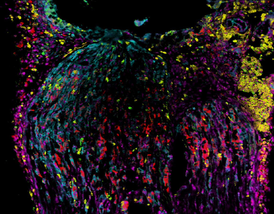
The cross-functional imaging innovation area comprises several imaging technologies that are used in research and diagnostics in the preclinical and clinical domains. These include classical histology and immunohistochemistry as well as state-of-the-art technologies such as non-invasive in vivo fluorescence and luminescence imaging in animal models, radiation-free examination of microcirculation in the hands (Xiralite platform) and High Content imaging for in vitro assays.

A core technology used is the »SMILE« (Sequential Multiple Immunohistochemistry for Localization- and Expression analysis) system for the multiple sequential immunohistochemistry and visualization of up to 100 proteins in the same sample. The images are subsequently analyzed to identify and quantify cellular microenvironments in a tissue as well as the effects of compounds on immune responses. This rapid and cost-effective multiplexing technique also allows for significant reduction in the amount of tissue isolated from biopsies and individualized therapy recommendations.


Core Competencies: 

  • SMILE System for multiplexing immunohistochemistry
  • In vivo fluorescence- and luminescence-imaging (IVIS Spectrum)
  • In vitro High-Content imaging using confocal fluorescence microscopy (e.g. ImageXpress)
  • Single cell calcium imaging
  • Radiation-free investigation of microcirculation of the hands (Xiralite)


Pathomechanisms and their mode of action

Bioinformatics based quantitative analysis of the cellular microenvironment is performed using the SMILE technology. This allows the detection and quantification of cellular microenvironments of immune cells and the prediction of functional interactions. Detailed mechanism of actions studies can be performed during drug treatment.

Pathomechanisms of chronic inflammatory skin diseases

Analysis of biopsies of patients with clinically confirmed hidradenitis suppurativa (acne inversa), psoriasis vulgaris and atopic dermatitis (neurodermatitis) are performed using the SMILE system and compared to skin from healthy subjects. As each sample undergoes multiplex analysis of up to 50 antibodies, it is possible to differentiate various immune and non-immune cells and characterize specific phenotypes (i.e. pro- vs. anti-inflammatory) are used. A bioinformatics based analysis allows the aims at the identification of characteristic cellular microenvironments, which in turn enable the identification of key mechanisms and target cells.

Early diagnosis of psoriasis arthritis

This project aims to identify biomarkers for the early diagnosis of psoriasis arthritis following characterization of plaques of patient biopsies. This makes use of the SMILE system with 50 antibodies allowing the differentiation of immune and non-immune cells as well as detection of specific phenotypes. This approach compares cellular microenvironments of biopsies from plaque psoriasis and psoriasis arthritis and allows the tracking of disease progression.

Pathogenesis and therapy of chronic polyneuropathies

Around 5 million people in Germany suffer from neuropathic pain, which develops after damage of neurons of the peripheral or central nervous system. These pain forms are very heterogeneous and are currently difficult to treat, since the available drugs are not very effective or are associated with significant side effects. Therefore the SMILE technology is employed to identify pathomechanisms, validate new targets as well as the mechanisms of action of experimental drugs for chemotherapy-, tumor-, trauma-, and diabetes-induced pain neuropathies. Here, the focus is analysis of endogenous signal mediators and their role in the development of acute and chronic neuropathies. 

Rimola V, Hahnefeld L, Zhao J, Jiang C, Angioni C, Schreiber Y, Osthues T, Pierre S, Geisslinger G, Ji RR, Scholich K, Sisignano M.
Lysophospholipids contribute to oxaliplatin-induced acute peripheral pain.
J Neurosci. 2020 Nov 4:JN-RM-1223-20
doi: 10.1523/JNEUROSCI.1223-20.2020


Cohnen J, Kornstädt L, Hahnefeld L, Ferreiros N, Pierre S, Koehl U, Deller T, Geisslinger G, Scholich K.
Tumors Provoke Inflammation and Perineural Microlesions at Adjacent Peripheral Nerves.
Cells. 2020 Jan 29;9(2):320
doi: 10.3390/cells9020320


Kern K, Schäfer SMG, Cohnen J, Pierre S, Osthues T, Tarighi N, Hohmann S, Ferreiros N, Brüne B, Weigert A, Geisslinger G, Sisignano M, Scholich K.
The G2A Receptor Controls Polarization of Macrophage by Determining Their Localization Within the Inflamed Tissue.
Front Immunol. 2018 Oct 1;9:2261
doi: 10.3389/fimmu.2018.02261


Treutlein EM, Kern K, Weigert A, Tarighi N, Schuh CD, Nüsing RM, Schreiber Y, Ferreirós N, Brüne B, Geisslinger G, Pierre S, Scholich K.
The prostaglandin E2 receptor EP3 controls CC-chemokine ligand 2-mediated neuropathic pain induced by mechanical nerve damage.
J Biol Chem. 2018 Jun 22;293(25):9685-9695
doi: 10.1074/jbc.RA118.002492


Pierre S, Linke B, Suo J, Tarighi N, Del Turco D, Thomas D, Ferreiros N, Stegner D, Frölich S, Sisignano M, Meyer Dos Santos S, deBruin N, Nüsing RM, Deller T, Nieswandt B, Geisslinger G, Scholich K.
GPVI and Thromboxane Receptor on Platelets Promote Proinflammatory Macrophage Phenotypes during Cutaneous Inflammation.
J Invest Dermatol. 2017 Mar;137(3):686-695
doi: 10.1016/j.jid.2016.09.036


Sisignano M, Angioni C, Park CK, Meyer Dos Santos S, Jordan H, Kuzikov M, Liu D, Zinn S, Hohman SW, Schreiber Y, Zimmer B, Schmidt M, Lu R, Suo J, Zhang DD, Schäfer SM, Hofmann M, Yekkirala AS, de Bruin N, Parnham MJ, Woolf CJ, Ji RR, Scholich K, Geisslinger G.
Targeting CYP2J to reduce paclitaxel-induced peripheral neuropathic pain.
Proc Natl Acad Sci U S A. 2016 Nov 1;113(44):12544-12549
doi: 10.1073/pnas.1613246113